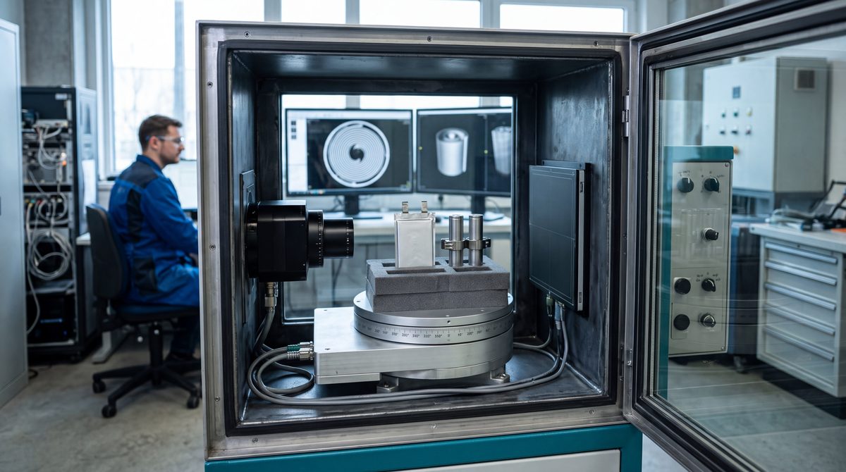
A batch of pouch cells comes through formation with no anomalies, then during aging three cells out of a few hundred show higher self-discharge than the rest. Opening them is the fastest way to find out why, and also the surest way to lose the evidence: cutting shifts the layers, the solvent evaporates, and the electrode that stuck out by a tenth of a millimeter is no longer where it was. With industrial CT the same cell can be examined layer by layer with the case still sealed, before anyone decides whether and where to section it.
For companies that make or assemble battery cells, modules and packs, in e-mobility as well as stationary storage, X-ray computed tomography has become an everyday development and quality tool. It is still a technique with hard physical limits. The page on industrial 3D CT and tomographic analysis covers the technique in general, while with lithium batteries what counts is the cell format, the detail you need to see and the number of samples, because resolution, scan time and the choice between an outside service and an in-house system depend on them.
Inside a cell, the defects that matter are tenths of a millimeter
In a lithium-ion cell the anode, usually graphite on copper foil, is slightly larger than the cathode, a metal oxide on aluminum foil. That margin is called overhang, and its job is to make sure every ion leaving the cathode finds graphite ready to take it. If the cathode reaches past the anode edge at some point, lithium can deposit as metal on that edge during charging, the so-called lithium plating, with capacity loss and in the worst cases dendrites growing through the separator. Design overhang is a fraction of a millimeter, and it has to be verified along the edges and above all at the corners, where stacking or winding is most likely to drift.
Most of the other defects CT looks for are born in production. Foreign metal particles, cutting debris and burrs on electrode edges can pierce the separator weeks later. In cylindrical cells the jelly roll, the wound stack of electrodes and separators, can deform toward the center, especially after many cycles. Tabs, welded by ultrasound or laser to the foils and terminals, can have incomplete welds or folds. Then come porosity and voids in the adhesives and thermal interface materials of modules, delamination of the active coating, and sealing defects, such as the heat seal of a pouch or the crimp of a cylindrical cell cap.
| Defect | Where it starts | What CT shows | Limit to keep in mind |
|---|---|---|---|
| Insufficient or negative anode-cathode overhang | Winding or stacking, electrode cutting | Distance between electrode edges, layer by layer, corners included | Needs a voxel of a few micrometers at the edge |
| Foreign particles | Slitting, notching, assembly environment | Metal inclusions denser than the active layers | Particles only a few voxels across, or made of light material, blend into noise |
| Jelly roll deformation | Winding, swelling during cycling | Collapsed or wavy layers, especially near the mandrel | A single scan cannot tell when the deformation began |
| Tab welds | Ultrasonic or laser welding | Position, folds, voids and separations in the joint | CT shows the joint geometry, its strength has to be tested by other methods |
| Porosity and voids | Adhesives, gap fillers, potting in modules | Distribution and volume of voids in the filler | The result depends on the segmentation threshold |
| Delamination | Calendering, cycling, internal gas | Separation between coating and current collector, swelling | Gaps thinner than the voxel stay invisible |
| Sealing | Pouch heat sealing, cap crimping | Continuity and geometry of the sealed area | A micro-leak of electrolyte calls for a leak test |
Why 2D radiography cannot measure overhang on its own
A radiograph projects the whole volume onto a plane, so dozens of electrode layers overlap in the same image. On a cylindrical cell, looking at the ends in profile, 2D radiography shows clearly whether the anode edges extend past the cathode edges, which is why many lines run it inline at full production rate. At the corners of a stacked cell, or on a large-format pouch, the overlap already makes it impossible to assign an edge to a specific layer.
Tomography rebuilds the volume from hundreds or thousands of projections taken at different angles, and lets you cut the cell virtually along any plane. That is how overhang is measured for every layer at all four corners, how a foil is followed along the whole winding, and how a particle is located in depth. The review by Pietsch and Wood in the Annual Review of Materials Research (2017) remains a good starting point on what tomography can reveal about electrodes and cells, from coating microstructure to the complete cell. The third dimension costs time, because a radiograph takes a fraction of a second and a high-resolution CT scan anywhere from many minutes to a few hours. That is why the two techniques work side by side in factories, with 2D inline on every cell and CT on samples and suspect parts.
Cell, module, pack: sample size sets the resolution
In cone beam CT the sample has to fit, entirely or almost, inside the field of view of the detector. The achievable voxel is therefore tied to the object diameter divided by the number of usable detector pixels. With a detector of about two thousand pixels, an object 20 mm wide is reconstructed with voxels on the order of 10 micrometers, one 200 mm wide on the order of 100. These are rough orders of magnitude, because focal spot, noise and artifacts make the real resolution worse than the nominal voxel. The guide ASTM E1441 and the practice ASTM E3375 describe these relationships and how system performance is verified. A common rule of thumb says a defect must span at least two or three voxels to be recognized reliably.
The most common cylindrical formats are 18 to 46 millimeters in diameter, and they are scanned whole with voxels from a few micrometers up to several tens. To resolve individual layers near the edges, a local high-magnification scan on a region of interest is often used, accepting some artifacts at the margins. Prismatic cells have a rigid aluminum or steel can and larger dimensions, which call for higher voltages and push the voxel toward tens of micrometers on the whole cell. Large-format pouches are the most awkward case, because they are flat and wide, and the beam crosses a lot of material when it travels in the plane of the cell. The usual approach is local scans of the corners, or laminography, a variant designed for flat objects.
On modules the scale changes completely. A whole module, with housing, cooling plates and dozens of cells, is reconstructed with voxels from a few tens to a few hundred micrometers. At that resolution overhang cannot be measured, while cell positions, busbar welds, adhesive continuity and voids in the thermal filler are checked well. A complete pack, short of dedicated installations, is handled as subassemblies. Scan times scale the same way, from a few minutes for a quick low-resolution scan to hours for a cell studied layer by layer.
Contrast depends on the materials. The copper of the anode collector and the nickel, manganese or cobalt oxides of the cathode attenuate far more than graphite and separator, which is why electrode edges stand out well. Lithium, a very light element, is almost transparent to X-rays. Lithium plating is inferred from its effects, such as swelling and separation, but it is rarely seen directly, and that is one reason researchers also use neutron tomography, as in the study by Ziesche and colleagues in Nature Communications (2020), which combines neutrons and X-rays on the same cell.
Want to look inside your cells before opening them?
Send us the cell format, chemistry, the defect you are looking for and the number of samples: we arrange a trial CT scan on your cells and tell you what resolution your format allows. Quote within 24 hours.
Email us Message us on WhatsAppFrom a fresh cell to a cycled one: what changes in the volume
One of the most useful applications belongs to development even before production. Scanning the same cell before and after a cycling campaign lets you compare two volumes and see where the structure has changed. In the work by Kok and colleagues in Sustainable Energy & Fuels (2019) a commercial cylindrical cell was scanned new and after 1,061 cycles, when its capacity had dropped to 79% of the initial value. The authors virtually "unrolled" the jelly roll to obtain a deformation map along the winding, and part of the capacity fade was attributed to delamination in the innermost layers.
Research has gone further, to watching cells while they fail. The group led by Finegan in Nature Communications (2015) used high-speed synchrotron tomography to follow thermal runaway in cylindrical cells, documenting gas formation, delamination and layer collapse. Such experiments need large research facilities and are out of reach for a quality lab. Their practical value lies in the catalog of mechanisms they made visible, which now guides how a post-mortem scan of a failed or field-returned cell is read.
For a failure analysis the typical sequence starts with radiography to get oriented, continues with a CT scan of the whole cell and then local scans of the suspect areas. Only at the end, if needed, is the sectioning plane chosen, with the CT volume pointing to where to cut. The destructive test comes last, when you already know what you are looking for and where.
Charged cells on the CT stage: handling rules
A lithium cell remains an energy source even inside a shielded cabinet, and a defective one more so than a healthy one. Before scanning, agree on the state of charge with whoever made the cell, often reduced for structural analysis, and protect the terminals with insulating tape or caps. Fixtures should be made of insulating, low-attenuation materials such as foams and engineering plastics, with no points that could crush or puncture the case. Swollen, dented or already failed cells are kept apart, in fire-resistant containers, and scanned with dedicated procedures.
Shipping to an outside lab follows strict rules as well. Lithium cells and batteries travel as dangerous goods, and for transport they must have passed the tests in section 38.3 of the UN Manual of Tests and Criteria. Prototypes and damaged or defective cells, which are often exactly the samples to be analyzed, fall under stricter special provisions on packaging and paperwork. It is wiser to hand shipping to a forwarder qualified for dangerous goods and to settle the number of samples before packing them. A non-disclosure agreement signed before shipment protects drawings, chemistry and process data.
Outside service or an in-house CT system
The choice depends mostly on frequency. An outside service makes sense for developing and qualifying new cells, for failure analysis, for qualifying a supplier, for first samples of a new module and in general when scans number a few dozen a year. It has a less obvious advantage too: it lets you tune the scan recipe, meaning voltage, filters, voxel, number of projections and analysis thresholds, before spending on a machine. The industrial CT scanning service is built for these cases, with a feasibility check on your format before the quote.
An in-house system starts to make sense when scans are daily, when production quality needs an answer within the same shift, or when samples cannot leave the plant. At that point the business case covers several items: cells scrapped or reworked without knowing why, hours of destructive teardown, the turnaround time of an outside lab, and the risk of a defect reaching the field. A CT scanner is, however, equipment that emits ionizing radiation, and in Italy it falls under Legislative Decree 101/2020, with the required notifications and the appointment of a radiation protection expert. It also takes floor space, trained operators and analysis software suited to battery measurements. To configure custom industrial CT systems, the safest route starts with a trial scan of the most demanding sample, the one that sets the voxel, and of the bulkiest one, which sets cabinet size and voltage.
Considering a CT system for your batteries?
Tell us your cell and module formats, the defects to check, the number of scans per day and whether you work in a lab or next to the line: we help you choose the CT system, starting from a trial scan of your samples. Quote within 24 hours.
Request a quote by email Message us on WhatsAppWhat battery standards require, and where CT fits in
Cell safety standards do not prescribe CT. IEC 62660-3 sets safety tests and acceptance criteria for lithium-ion cells used to propel electric vehicles, while IEC 62619 covers cells and batteries for industrial applications, energy storage included. Both check behavior under reasonably foreseeable misuse, and an internal defect such as a particle or a negative overhang is exactly one of the ways an apparently healthy cell can end up in a short circuit. Tomography is how you show, before testing and after, that the cell is built as designed.
On the regulatory side, Regulation (EU) 2023/1542 on batteries introduces safety requirements for stationary energy storage systems, information duties and, from 2027, a digital passport for electric vehicle batteries, light means of transport batteries and industrial batteries above 2 kWh. No article mandates CT, yet a quality system that documents the internal build of cells, batch by batch, already holds much of the data customers and authorities may ask for. For an overview of industrial CT applications, from metrology to defects, the review by De Chiffre and colleagues in CIRP Annals (2014) is still useful.
In practice you start from a real cell in the beam and a precise question, for example what minimum overhang must be guaranteed or which particle is already too large. With that question and the sample format, a trial scan shows whether CT gives the answer, at what voxel and in how much time, and those numbers decide between service and system.
What to send us for an assessment
To propose a trial scan or a suitable system we only need a few details:
- format and dimensions of the cell, module or pack (cylindrical, prismatic, pouch);
- chemistry and case materials, if you can share them;
- the defect to find or the measurement to make (overhang, particles, tab welds, voids in the gap filler);
- the smallest detail to detect, even a rough figure;
- number of samples, state of charge and condition (new, cycled, failed);
- expected scans per month and whether you are thinking of a service or an in-house system.
You can send them by email to info@dipitech.com or on WhatsApp. You can also use the contact form.
With these details we arrange a trial CT scan of your cells through the outside CT analysis service, or we help you choose a custom CT system for cells and modules, with a quote within 24 hours and technical support in Italy. Other applications are described on the industrial CT page.
Request a quote
Leave your name, email, and a few lines about your application, with no obligation. We reply within 24 hours with initial technical guidance and a quote.
Frequently asked questions about CT inspection of lithium batteries
What is industrial CT and what are its advantages for batteries?
It is an X-ray technique that rebuilds the internal volume of an object from many projections taken at different angles. On cells and modules it measures anode-cathode overhang, finds foreign particles and assesses winding deformation, tab welds and voids in adhesives without opening the case, so the evidence is not destroyed before you know where to look.
What resolution is needed to measure cell overhang?
To resolve the edges of individual electrodes you need voxels of a few micrometers at the edge. On cylindrical cells this is reached on the whole cell or with local high-magnification scans; on large-format pouches the corners are handled with local scans or laminography. On whole modules the voxel rises to tens or hundreds of micrometers and overhang cannot be measured.
How long does a CT scan of a battery cell take?
It depends on the required resolution and the format. A quick low-resolution scan takes a few minutes, a high-resolution scan to study the cell layer by layer anywhere from many minutes to a few hours. That is why production lines run 2D radiography on every cell and CT on samples and suspect parts.
Can X-ray CT see lithium plating?
Rarely directly, because lithium is a light element and almost transparent to X-rays. Plating is inferred from its effects, such as swelling, separation and layer deformation. To observe lithium itself, researchers also use neutron tomography, often combined with X-ray CT.
How are cells shipped for an outside CT scan?
Lithium cells and batteries travel as dangerous goods and must have passed the tests in section 38.3 of the UN manual; prototypes and damaged cells follow stricter special provisions. Use a forwarder qualified for dangerous goods, agree on the state of charge, insulate the terminals and sign a non-disclosure agreement before shipping.
Outside CT scanning service or an in-house CT system?
A service suits development, failure analysis, supplier qualification and workloads of a few dozen scans a year. An in-house system makes sense with daily scans, answers needed within the same shift or samples that cannot leave the plant, keeping radiation protection requirements in mind. DIPITECH arranges a trial CT scan of your cells and helps you choose, with a quote within 24 hours.